-
Home
-
About JCTR
-
Gold Open Access
-
Issues
-
Editorial board
-
Author guidelines
-
Publication fees
-
Online first
-
Special issues
-
News
-
Publication ethics
-
Partners
-
Submit your manuscript
-
Submit your review report
-
Editorial Office
-

This work is licensed under a Creative Commons Attribution-NonCommercial 4.0 International License. ISSN print: 2382-6533 ISSN online: 2424-810X
Volume 8 Issue 6
Interobserver variability in GTV contouring in non-spine bone metastases
Carolina de la Pinta*, Raquel García LaTorre, Alberto Martínez-Lorca, Eva Fernández, Raul Hernanz, Mercedes Martín, Jose A. Domínguez, Teresa Muñóz, Elena Canales, Carmen Vallejo, Marina Alarza, Asunción Hervás, Manuel Garví, Vanesa Pino, Sonsoles Sancho
de la Pinta et al. J Clin Transl Res 2022; 8(5):3
Published online: October 22, 2022
Abstract
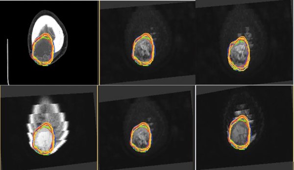
Background and aim: The optimal imaging test for GTV (gross tumor volume) delineation in non-spine bone metastases has not been defined. The use of stereotactic body radiotherapy requires accurate target delineation. MRI and/or 18FDG-PET allows for better visualization of the extent of bone metastases and optimizes the accuracy of tumor delineation for stereotactic radiotherapy compared to CT alone. We evaluated the interobserver agreement in GTV of non-spine bone metastases in a single center and compared MRI and/or 18FDG-PET and CT in GTV delineation.
Methods: Anonymous CT and MRI and/or 18FDG-PET obtained from 10 non-spine bone metastases were analyzed by 6 radiation oncologists at our center. Images acquired by CT and MRI and/or 18FDG-PET were used to delineate 10 GTVs of non-spine bone metastases in the pelvis, extremities, and skull. The cases showed different characteristics: blastic and lytic metastases, and different primary cancers (lung, breast, prostate, rectum, urothelial, and biliary). In both CT and MRI and/or 18FDG-PET, the GTV volumes were compared. The index of agreement was evaluated according to Landis and Koch protocol.
Results: The GTV volume as defined on MRI was in all cases larger or at least as large as the GTV volume on CT (p = 0.25). The median GTV volume on MRI was 3.15 cc (0.027–70.64 cc) compared to 2.8 cc on CT (0.075–77.95 cc). Interobserver variance and standard deviation were lower in CT than MRI (576.3 vs 722.2, and 24.0 vs 26.9, respectively). The level of agreement was fair (kappa = 0.36) between CT and MRI. The median GTV volume on 18FDG-PET in five patients was 5.8 cc (0.46–64.17 cc), compared to 4.1 cc on CT (0.99–54.2 cc) (p = 0.236). Interobserver variance and standard deviation in CT, MRI, and 18FDG-PET were 576.3 vs 722.2 vs 730.5 and 24 vs 26.9 vs 27.0, respectively. The level of agreement was slight (kappa = 0.08) between CT and 18FDG-PET.
Conclusions: Interobserver variance in non-spine bone metastases was equal when MRI and PET were compared to CT. CT was associated with the lowest variance and standard deviation. Compared to CT GTVs, the GTVs rendered from MRI images had fair agreement while the GTVs rendered from 18FDG-PET had only slight agreement.
Relevance for patients: The delimitation of the treatment volume in non-spine bone metastases with SBRT is important for the results determining its efficacy. It is therefore essential to know the variability and to manage it in order to achieve the highest quality of treatment.
DOI:http://dx.doi.org/10.18053/jctres.08.202206.003
Author affiliation
1. Radiation Oncology Department, Ramón y Cajal University Hospital, IRYCIS, Madrid, Spain
2. Radiology Department, Ramón y Cajal University Hospital, IRYCIS, Madrid, Spain
3. Nuclear Medicine Department, Ramón y Cajal University Hospital, IRYCIS, Madrid, Spain
*Corresponding author
Carolina de la Pinta
Radiation Oncology Department. Ramón y Cajal University Hospital, IRYCIS, Alcalá University, 28034
Madrid, Spain.
E-mail: cdelapinta88@gmail.com
Handling editor:
Michal Heger
Department of Pharmaceutics, Utrecht University, the Netherlands
Department of Pharmaceutics, Jiaxing University Medical College, Zhejiang, China