-
Home
-
About JCTR
-
Gold Open Access
-
Issues
-
Editorial board
-
Author guidelines
-
Publication fees
-
Online first
-
Special issues
-
News
-
Publication ethics
-
Partners
-
Submit your manuscript
-
Submit your review report
-
Editorial Office
-

This work is licensed under a Creative Commons Attribution-NonCommercial 4.0 International License. ISSN print: 2382-6533 ISSN online: 2424-810X
Volume 7 issue 4
Development of a new decellularization protocol for the whole porcine heart
Ana Lídia Jacintho Delgado, Ana Claudia Oliveira Carreira, Hianka Jasmyne Costa de Carvalho, Renata Kelly da Palma, Taís Harumi de Castro Sasahara, Carla Maria Figueiredo de Carvalho, Marisol León, Rodrigo da Silva Nunes Barreto, Maria Angélica Miglino*
Delgado et al. J Clin Transl Res 2021; 7(4):17
Published online: August 8, 2021
Abstract
Background: Cardiovascular diseases are the leading cause of death in many countries. Advances in technology have been promoted in this regard, especially in Tissue Engineering, to meet the need for tissue or organ grafts. In this way, the porcine model has been used due to his morphophysiological similarity between the human species, mainly regarding the cardiovascular system. Tissue engineering is employed using biological scaffolds that are currently derived from porcine. These scaffolds are produced by decellularization, a process to remove cells aiming to maintain only its three-dimensional structure, formed by extracellular matrix. Its main objective is to produce organs with recellularized scaffolds that could eventually substitute the ones with impaired functions.
Aim: In this way, the present study aimed to establish a new protocol for porcine heart decellularization with potential application on tissue engineering.
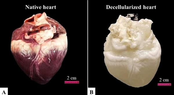
Methods: A porcine heart aorta was cannulated with a silicon tube, and the organ was washed in phosphate-saline buffer 0.1% solution through a peristaltic pump (Harvard Peristaltic Pump - Harvard Apparatus). After that, deionized water was perfused in the same system. The decellularization was carried out by perfusing four liters of 4% Sodium Dodecyl Sulfate were through myocardial circulation, at 400 mL / min for 24 hours, for 6 days; perfusion of Triton X-100 1% followed by phosphate saline buffer 0.1M and deionized water for 24 hours. The heart volume was measured before and after the recellularization. Macroscopic and microscopic analyses were performed on the cardiac tissue, and Hematoxylin & Eosin, Masson’s Trichrome, Weigert-Van Giesson, Alcian Blue and Picrosirius Red staining were evaluated. To observe the cell adhesion, the recellularized was provided in this scaffold, which was analyzed under Immunofluorescence and Scanning Electronic Microscopy.
Results: The protocol provided cells remotion, with adequate concentration of remaining DNA. Extracellular matrix components as collagen type I, elastin, glycosaminoglycans were successfully maintained. The scaffold showed a high cells adherence and proliferation in the recellularization process.
Conclusions: According to results, the protocol described in this work preserved the extracellular matrix and architecture of the organ, minimizing extracellular matrix loss and being possible to state that it is a promising approach to tissue bioengineering.
Relevance for patients: This study provides a protocol for whole porcine heart decellularization, which will ultimately contribute to heart bioengineering and may support further studies on biocompatibility relationship of new cells with recellularized scaffolds.
DOI: http://dx.doi.org/10.18053/jctres.07.202104.017
Author affiliation
Department of Surgery, School of Veterinary Medicine and Animal Science, University of São Paulo, São Paulo, Brazil
*Corresponding author
Maria Angélica Miglino
Department of Surgery, School of Veterinary Medicine and Animal Science, University of São Paulo, Avenue Prof. Dr. Orlando Marques de Paiva, 87, Cidade Universitária, Butantã, São Paulo, SP, CEP: 05508-270, Brazil.
Tel: +55 (11) 30917690
Email: miglino@usp.br
Handling editor:
Michal Heger
Department of Pharmaceutics, Utrecht University, the Netherlands
Department of Pharmaceutics, Jiaxing University Medical College, Zhejiang, China