-
Home
-
About JCTR
-
Gold Open Access
-
Issues
-
Editorial board
-
Author guidelines
-
Publication fees
-
Online first
-
Special issues
-
News
-
Publication ethics
-
Partners
-
Submit your manuscript
-
Submit your review report
-
Editorial Office
-

This work is licensed under a Creative Commons Attribution-NonCommercial 4.0 International License. ISSN print: 2382-6533 ISSN online: 2424-810X
Volume 4 Issue 2
Feasibility of mapping and cannulation of the porcine epicardial lymphatic system for sampling and decompression in heart failure research
Benjamin Kappler, Dara R. Pabittei, Sjoerd van Tuijl, Marco Stijnen, Bas A.J.M. de Mol, Allard C. van der Wal
Kappler et al., J Clin Tranl Res 2018; 4(2): 2
Published online: July 18, 2018
Abstract
Background and Aim: The cardiac lymphatic system drains excess fluid from the cardiac interstitium. Any impairment or dysfunction of the lymph structures can result in the accumulation of interstitial fluid, and may lead to edema and eventually cardiac dysfunction. Lymph originates directly from the interstitium and carries real-time information about the metabolic state of cells in specific regions of the heart. The detailed anatomy of the epicardial lymphatic system in individuals is broadly unknown. Generally, the epicardial lymphatic system is not taken into consideration during heart surgery. This study investigates the feasibility of detailed mapping and cannulation of the porcine epicardial lymphatic system for use in preservation of explanted hearts and heart failure studies in pigs and humans.
Methods: The anatomy of the epicardial lymphatic systems of forty pig hearts was studied and documented. Using a 27 G needle, India ink was introduced directly into the epicardial lymphatic vessels in order to visualise them. Based on the anatomical findings thus obtained, two cannulation regions for the left and right principal trunks were identified. These regions were cannulated with a 26 G intravenous Venflon cannula-over-needle, and a Galeo Hydro Guide F014 wire was used to verify that the lumen was patent.
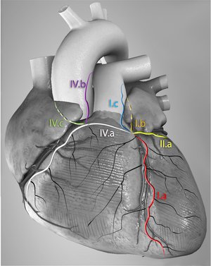
Results: The main epicardial lymphatic collectors were found to follow the main coronary arteries. Most of the lymph vessels drained into the left ventricular trunk, which evacuates fluid from the left heart and also partially from the right heart. The right trunk was often found to drain into the left trunk anterior basally. Right heart drainage was highly variable compared to the left. In addition, the overall cannulation success rate of the selected cannu-lation sites was only 57%.
Conclusions: Mapping of the porcine epicardial lymphatic anatomy is feasible. The right ventricular drainage system had a higher degree of variability than the left, and the right cardiac lymph system was found to be partially cleared through the left lymphatic trunk. To improve cannulation success rate, we proposed two sites for cannulation based on these findings and the use of Venflon cannulas (26 G) for cannulation and lymph collection. This method might be helpful for future studies that focus on biochemical sample analysis and decompression.
Relevance for patients: Real-time biochemical assessment and decompression of lymph may contribute to the understanding of heart failure and eventually result in preventive measures. First its relevance should be established by additional research in both arrested and working porcine hearts. Imaging and mapping of the epicardial lymphatics may enable sampling and drainage and contribute to the prevention or treatment of heart failure. We envision that this approach may be considered in patients with a high risk of postoperative left and right heart failure during open-heart surgery.
 Anterior view of the principal routes of the porcine epicardial lymphatic drainage
DOI: http://dx.doi.org/10.18053/jctres.04.201802.002
Author affiliation
1 LifeTec Group B.V., Eindhoven, The Netherlands
2 Academic Medical Center, Department of Cardiothoracic Surgery, Amsterdam, The Netherlands
3 Hasanuddin University Faculty of Medicine, Department of Physiology, Makassar, Indonesia
4 Academic Medical Center, Department of Pathology, Amsterdam, The Netherlands
*Corresponding author
Benjamin Kappler
LifeTec Group B.V., 10-11, Kennedyplein, 5611 ZS Eindhoven, The Netherlands
Tel: +31 40 298 9393
Email: b.kappler@lifetecgroup.com
Handling editor:
Michal Heger
Department of Experimental Surgery, Academic Medical Center, University of Amsterdam, Amsterdam, the Netherlands

